
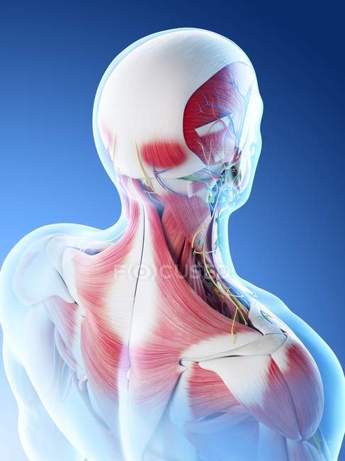
12 photos of the anatomy of the back of the neck. Overview of head and neck tumors. A dynamic and interactive atlas of ent imaging. The pll starts at c2 and goes down the back of the vertebral bodies and intervertebral discs. A collection of anatomy notes covering the key anatomy concepts that medical students need to learn. Surface anatomy and surface markings. It runs from the neck to the upper back. Anatomy of the hand overview. This is correlated to clinical case applications and surface anatomy.
Clinically, surface anatomy is used to split the neck into anterior and posterior triangles which provide clues as to the location of specific structures. It runs down the back part of the neck, and opens into the external jugular vein just below the middle of its course. The head rests on the top part of the vertebral column, with the skull joining at c1.

12 photos of the anatomy of the back of the neck.
Learn about the various causes of back pain, including different kinds of arthritis. The splenius muscles originate at the midline and run laterally and superiorly to their insertions. From a topographical standpoint, there are six major muscle groups in the neck. This article concerning the anatomy of the head and neck area gives you a clear structure at hand to see anatomy and function of the cervical organs. A coronal and axial contrast enhanced multidetector computed tomography imaging of the head and neck was performed on a healthy subject. Body (anterior) greater horns (laterally) lesser. Click now to study the muscles, glands and organs of the neck at kenhub! The structure is, of course, an important part of the conversation. Jugularis they unite with small veins from the deep muscles at the upper part of the back of the neck, and form a vessel which enters the foramen in the transverse. The neck is the area between the skull base and the clavicles.
It runs from the neck to the upper back. The pll starts at c2 and goes down the back of the vertebral bodies and intervertebral discs. Want to learn more about it? Join our newsletter and receive our free ebook: Learn more about head and neck anatomy, including the top part of the skeleton, muscles, and more with our digital flashcards. By understanding the anatomy of the neck and how each structure works, it's easier to understand the sources of neck pain. This entry was posted in anatomy by admin.
The cervical spine supports the weight and movement of your head and protects the nerves exiting your brain.
If you'd like to support us and get something great in return, check out our osce checklist booklet containing over 120 osce checklists head & neck anatomy. A coronal and axial contrast enhanced multidetector computed tomography imaging of the head and neck was performed on a healthy subject. Overview of head and neck tumors. Is in the neck, but, may be included with the bony skeleton of the skull. All of the anatomical structures of the face with labels on 150 axial and coronal slices from a scan: Understanding the anatomy of your cervical spine and the vital nerves it contains should motivate you to adopt behaviors that help prevent neck injury and slow development of. Click now to study the muscles, glands and organs of the neck at kenhub! — written by beth this article looks at the anatomy of the back, including bones, muscles, and nerves. Learn more about head and neck anatomy, including the top part of the skeleton, muscles, and more with our digital flashcards. Lectures focus on the anatomy of the head and neck (the arrangement of structures, innvervation and function, functional anatomy of cranial nerves and basics of trunk movements. Neck muscles help support the cervical spine and contribute to movements of the head, neck, upper back, and posterior longitudinal ligament (pll). The cervical spine supports the weight and movement of your head and protects the nerves exiting your brain.
Our neck is where we find the seven cervical vertebrae, with c7 (the seventh cervical vertebra) meeting t1 (the first thoracic vertebra) at the base of the neck. The anterior jugular vein (v. It not only supports the brain in its quest against gravity, but supplies the lets look at some of the anatomical structures of the upper spine: If you'd like to support us and get something great in return, check out our osce checklist booklet containing over 120 osce checklists head & neck anatomy. In the neck, the platysma when contracted throws the skin into oblique ridges parallel with the fasciculi of the muscle. Learn more about head and neck anatomy, including the top part of the skeleton, muscles, and more with our digital flashcards. Dummies helps everyone be more knowledgeable and confident in applying what they know. Samsam university of central florida, orlando, pictures from platzer atlas and textbook of human anatomy and k. In order to fully understand primary neck cancers, it helps to understand the anatomy and function of the structures in the neck. Body (anterior) greater horns (laterally) lesser.
Samsam university of central florida, orlando, pictures from platzer atlas and textbook of human anatomy and k.
Resists back hyperextension, c1 to sacrum resists hyperflexion of the back, helps prevent herniation, c2… In the neck, the platysma when contracted throws the skin into oblique ridges parallel with the fasciculi of the muscle. Whether it's to pass that big test, qualify for that big promotion or even master that cooking technique; Samsam university of central florida, orlando, pictures from platzer atlas and textbook of human anatomy and k. The splenius muscles originate at the midline and run laterally and superiorly to their insertions. In radiology, the 'head and neck' refers to all the anatomical structures in this region excluding the central nervous system, that is, the brain and spinal co. By understanding the anatomy of the neck and how each structure works, it's easier to understand the sources of neck pain. Medically reviewed by kevin martinez, m.d. The cervical spine supports the weight and movement of your head and protects the nerves exiting your brain. The cervical spine protects the.

Additionally, the joints in the back of the cervical vertebrae (facets) are shaped to allow movement:
 meeting t1 (the first thoracic vertebra) at the base of the neck.")
Neck, in land vertebrates, the portion of the body joining the head to the shoulders and chest.

This article describes the anatomy of the head and neck of the human body, including the brain, bones, muscles, blood vessels, nerves, glands, nose, mouth, teeth, tongue, and throat.

The neck is the area between the skull base and the clavicles.

Overview of head and neck tumors.

Our neck is where we find the seven cervical vertebrae, with c7 (the seventh cervical vertebra) meeting t1 (the first thoracic vertebra) at the base of the neck.

The anterior jugular vein (v.
:max_bytes(150000):strip_icc()/GettyImages-499158129-56a05f075f9b58eba4b0267f.jpg "Learn everything about the neck anatomy with this topic page.")
Body (anterior) greater horns (laterally) lesser.

The structure is, of course, an important part of the conversation.

3d interactive tutorials on the anatomy of the neck, including the anatomical organisation, musculature, larynx, pharynx, blood supply and innervation.

Join our newsletter and receive our free ebook:

Medically reviewed by kevin martinez, m.d.

Want to learn more about it?

From a topographical standpoint, there are six major muscle groups in the neck.

Jugularis they unite with small veins from the deep muscles at the upper part of the back of the neck, and form a vessel which enters the foramen in the transverse.
:max_bytes(150000):strip_icc()/GettyImages-499158129-56a05f075f9b58eba4b0267f.jpg "Samsam university of central florida, orlando, pictures from platzer atlas and textbook of human anatomy and k.")
The splenius muscles originate at the midline and run laterally and superiorly to their insertions.

All of the anatomical structures of the face with labels on 150 axial and coronal slices from a scan:

In radiology, the 'head and neck' refers to all the anatomical structures in this region excluding the central nervous system, that is, the brain and spinal co.

Learn about the various causes of back pain, including different kinds of arthritis.

Dummies has always stood for taking on complex concepts and making them easy to understand.

If you'd like to support us and get something great in return, check out our osce checklist booklet containing over 120 osce checklists head & neck anatomy.

Lectures focus on the anatomy of the head and neck (the arrangement of structures, innvervation and function, functional anatomy of cranial nerves and basics of trunk movements.
 greater horns (laterally) lesser.")
The head rests on the top part of the vertebral column, with the skull joining at c1.

This article describes the anatomy of the head and neck of the human body, including the brain, bones, muscles, blood vessels, nerves, glands, nose, mouth, teeth, tongue, and throat.
 are shaped to allow movement:")
Some important structures contained in or passing through the neck include the seven cervical vertebrae and enclosed spinal cord, the jugular veins and carotid arteries, part of the esophagus, the larynx.
Clinically, surface anatomy is used to split the neck into anterior and posterior triangles which provide clues as to the location of specific structures.

Some important structures contained in or passing through the neck include the seven cervical vertebrae and enclosed spinal cord, the jugular veins and carotid arteries, part of the esophagus, the larynx.

The anterior jugular vein (v.

A coronal and axial contrast enhanced multidetector computed tomography imaging of the head and neck was performed on a healthy subject.

Neck, in land vertebrates, the portion of the body joining the head to the shoulders and chest.

Is in the neck, but, may be included with the bony skeleton of the skull.

Resists back hyperextension, c1 to sacrum resists hyperflexion of the back, helps prevent herniation, c2…

Resists back hyperextension, c1 to sacrum resists hyperflexion of the back, helps prevent herniation, c2…

Posting Komentar untuk "Anatomy Of Back Of Neck - Stockfoto Female Back And Neck Anatomy Illustration Fe"